Craig Barrington, DDS, knows how to make teeth transparent. It seems implausible, but with enough know-how, you can diaphanize the hard enamel and dentin structures of extracted teeth to literally see what lies inside. It turns out, there is a lot to see.
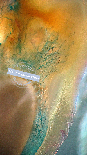
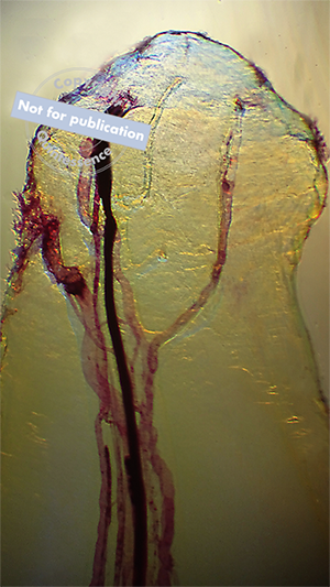
“The dental pulp is an amazing and complex structure.” Dr Barrington continues, “There is not a lot of information available on the vascularity and nerve distribution within a human tooth. Most—if not all—pictures of the vascularity and innervation of the human pulp are artist-generated depictions of what they ‘believe’ or even ‘wish’ is inside our teeth. There is certainly more than we think hiding inside each and every single tooth.”
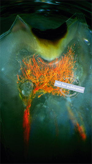
Like most people, I remember the first time I saw one of Dr Barrington’s images. The photograph showed the root of a tooth, but somehow, impossibly, the tooth structure was transparent, and the pulp canals were clearly visible. It was beautiful and arresting, and I had never seen anything like it. Even though I was familiar with his work, the collection of images in his new book, An Atlas of Dental Vascularity & Innervation, far surpasses anything I could have imagined. Throughout its pages, Dr Barrington shows us the beauty and grandeur of the dental pulp. The images speak for themselves, and they answer important questions about how nerves and blood vessels are distributed within teeth and the kinds of variation that exist for internal dental anatomy.
“We are missing a lot in dentistry. We extract and discard human teeth like they are trash. No other field of medicine trashes and discards human body parts at the same rate and with the same disregard as found in the field of dentistry. It would be my hope that the anatomical images featured in these pages would change this and set us on a new path.”
Preview Dr Barrington’s book here.
 Craig Barrington, DDS, graduated summa cum laude in 1996 from the School of Dentistry at the University of Texas Health Science Center at San Antonio. Microscopes and histology have always been an interest of his, but the first time he looked at a transparent human tooth, he knew that this would be a lifelong focus of his research. To this end, Dr Barrington has made it his mission to understand every diaphanization method that he has come across. He has learned how, where, and why transparency can occur in a solid object. Dr Barrington holds a patent in histology and multiple copyrights. He maintains a private practice in Waxahachie, Texas.
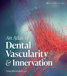 An Atlas of Human Dental Vascularity & Innervation
Craig Barrington
Our understanding of internal dental anatomy has remained limited by our inability to see inside a tooth without sectioning it. However, for one dentist working to find a way to see inside a tooth, the answer was diaphanization. This atlas represents the breathtaking results of photographing human teeth that have been made transparent. Dr Barrington has learned as many diaphanization methods as possible to understand how, where, and why transparency can occur in a solid object and translated that knowledge to transforming tooth structure. The images in this atlas showcase the internal anatomy of the teeth, with a special emphasis on the innervation and vascular structure and their distribution within the dentin chamber. For each image, the author follows a complex diaphanization method to make an extracted tooth transparent, before photographing the intact internal dental anatomy. Therefore, the images in this book display structures that have rarely been seen so clearly and in three dimensions, including the pulp chamber, apical anatomy, tooth channels, as well as pulpal pathology. This atlas pushes our understanding of internal dental anatomy and serves as an inspiration as to what one individual can do to advance knowledge within dentistry.
144 pp; 178 illus; ©2022; ISBN 978-1-64724-100-1 (B1001); US $104 (preorder)