“Kids these days!”
 What comes to mind when you hear that phrase? Young people constantly on their phone instead of reading books or newspapers? Someone seeing a piece of tech from the 20th century and ignorantly asking “what’s that?” A youngster fresh out of school who thinks he knows better than those who have been practicing longer than he’s been alive, just because he grew up with the Internet and is well-versed on the most cutting-edge innovations?
A common theme among these examples is the role of technology in the separation of the generations. No matter how old you are, there are some products, ideas, and practices that were already outdated when you were entering the workforce, some that became established as you were becoming established as a professional, and some that you’ve had to teach yourself to adapt to in order to continue providing the highest quality of care. Of course, even the most seasoned dentists have a responsibility to keep up with newer technology, especially as it has been proven to improve patient outcomes, but what about teaching older methods to the younger generation?
Even when there are new options available, the learner is lacking if they remain ignorant of the materials and methods that came before them—especially when these methods build the foundation for those that come next. Just because we all carry around calculators in our pockets, it doesn’t mean we shouldn’t know how to add, and even if our devices come with a spell check, we should still know how to spell.
These were the principles at play in the decision to pursue a brand-new edition of Johan Reyneke’s timeless text, Essentials of Orthognathic Surgery. As Prof Reyneke explains, since the orthognathic surgery “renaissance” in the 1970s, our knowledge and understanding of all aspects of orthognathic surgery has increased greatly. However, a thorough understanding of the basic principles of diagnosis and treatment planning, including the development of a cephalometric visual treatment planning and model surgery, still forms the cornerstones of successful treatment. Many years ago, Auguste Comte (1798–1857) said, “One does not know a science completely without knowing its history.”
This is very true. But this book goes far beyond the history of orthognathic surgery. It’s all about the “essentials”—everything a burgeoning oral surgeon needs to know to treatment plan and carry out both basic and complex surgeries involving the lower third of the face.

Drawing and tracing to plan surgical treatment for different situations.
More than two thousand years ago, Cicero (106–143 BCE) recognized the importance of the human face when he wrote, “Everything is in the face … ”. The morphology of the human face consists of relatively few objects: the forehead, two eyes, two cheeks, a nose, and a chin; however, the unique shape, size, and relationships between these objects makes individual facial features recognizable among literally billions of individuals. The face of an individual not only reveals their identity and origin, but a person’s emotions, such as happiness, sadness, fear, anger, and traits such as friendliness. Facial esthetics is often subconsciously associated with character or personality.

Before her orthodontic treatment and orthognathic surgery, this girl was often bullied by her peers about the appearance of her face and jaw.
As Prof Reyneke says, “We don’t just change faces; we change lives.” The stakes are overwhelmingly high, and the clinician must do everything in their power to get it right. Currently, we are privileged to have access to exquisite CT studies with 3D data from our patients, and the surgeon may be tempted to leave the clinical evaluation and treatment planning to the available virtual treatment programs. The computer and technician, however, diagnose by “the numbers.” Farhad Naini in 2011 wrote wisely, “Nowhere in the field of medicine is the fusion of art and science more important than in the clinical assessment of facial esthetics.” As Prof Reyneke explains, “I am convinced that it is essential for the orthodontist and orthognathic surgeon to acquire a basic knowledge of clinical assessment and data analysis and also to develop an artistic eye when diagnosing and planning their patient’s treatment. We should use all the available data obtained from our clinical assessment as definitive diagnostic tool.” In the third edition of Essentials in Orthognathic Surgery, the essential basic principles are used in combination with advanced virtual treatment planning. The reader is taught how to evaluate and analyze the face shapes and landmarks, and how to plan dozens of types of surgery with prediction tracings. Case after case of striking before-and-after comparisons demonstrates the results that can be achieved when these principles are mastered.

Case example of a patient with hemifacial microsomia.
Virtual 3D planning is an incredible tool. There is no doubt that hardware and software will only continue to evolve with greater precision and clarity. Knowing how to use these technologies to their greatest advantage will just become more and more a requisite skill. But they are no substitute for the fundamental knowledge of how a face should look and how the bones and muscles will move during a surgery. There is a reason the first two editions of Essentials have become a favorite reference for orthodontic and surgical residents in training, as well as for practicing oral and maxillofacial surgeons. This third edition will only continue that trend.
Preview the book here.
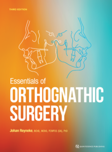 Essentials of Orthognathic Surgery
Johan Reyneke
This long-awaited new edition of the author’s seminal text on orthognathic surgery includes not only a fresh new look and over a dozen new cases, but essential updates for anyone practicing orthognathic surgery. Though many of the surgical practices and techniques have not drastically changed since the previous edition, recent research has inspired new sections on airway management and orthognathic surgery of the temporomandibular joint. The previous chapter on treatment of dentofacial deformities has now been split into two streamlined chapters on typical and complex dentofacial deformities to accommodate new cases and information, allowing a more user-friendly experience for the reader. Those familiar with the second edition will remember the illustrated step-by-step processes for patient evaluation, diagnosis, treatment planning, and surgical technique, and these vital resources have made it to the new text as well. From the most basic bilateral sagittal split osteotomy to complex surgery involving three-dimensional analysis, movement, and rotation of both jaws, this book will help everyone from surgical residents to experienced clinicians in managing both children and adults with dentofacial deformities, improving both function and esthetics.
 Johan P. Reyneke, BChD, MChD, FCMFOS (SA), PhD, is currently the director of the Centre for Orthognathic Surgery in Cape Town, South Africa, Visiting Professor at the University of Amsterdam, and an associate professor in the Division of Oral and Maxillofacial Surgery at the School of Dentistry at the Universidad Autónoma de Nuevo León in Monterrey, Mexico. He holds clinical professorships in the departments of oral and maxillofacial surgery at both the University of Florida in Gainesville, Florida, and the University of Oklahoma Health Sciences Center in Oklahoma City, Oklahoma. Dr Reyneke lectures extensively and has been an invited guest speaker at conferences on five continents. He has published several manuals on surgical technique, written chapters in several oral and maxillofacial surgery textbooks, has authored more than 65 peer-reviewed articles, and is coauthor of the book Introduction to Orthognathic Surgery: A Color Atlas (Ishiyaku EuroAmerica, 1991). Dr Reyneke has served on the national executive committee of the South African Society of Maxillofacial and Oral Surgeons for 14 years and was president for 4 years. He maintained a private practice in Johannesburg, South Africa, for four decades.
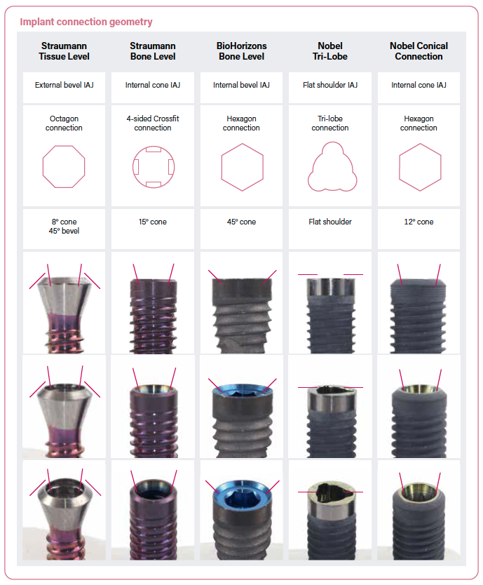
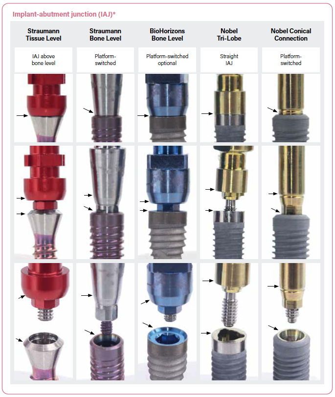
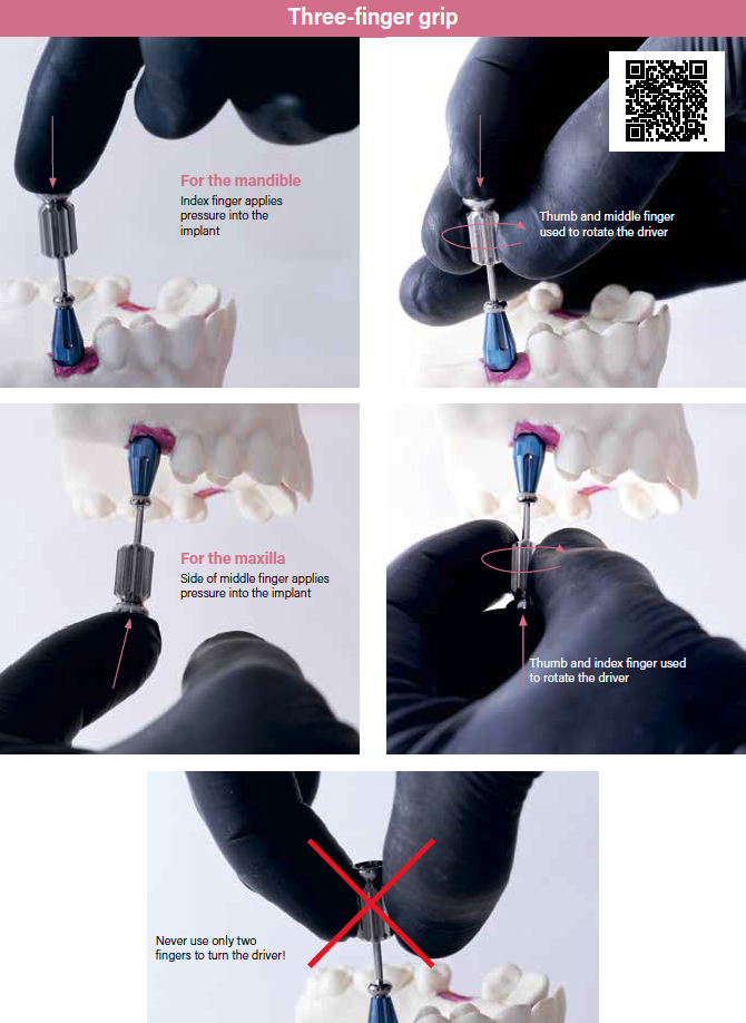
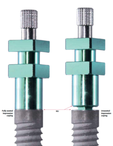 This is just one example of many where he takes the time to explain how to perform individual steps properly to maximize patient safety and clinical success. He is also wise to show what things should and shouldn’t look like at any given step. Take impression copings, for example. This is what proper and improper seating look like.
 Todd R. Schoenbaum, DDS, MS, is a Professor at the Dental College of Georgia, where he serves as the Coordinator for Implant Education and Related Research, training residents and students in implant restorations and clinical research. He was previously full Clinical Professor at the University of California, Los Angeles (UCLA) and Director of UCLA Continuing Education. Dr Schoenbaum has published over 50 papers and 2 textbooks, and he is the recipient of the scientific writing award from the Journal of Prosthetic Dentistry. He has a master’s in clinical research from the UCLA School of Medicine, and he has been invited to present his clinical and scientific work at conferences worldwide.
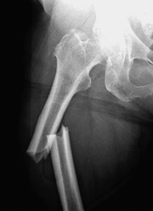 Take a look at this radiograph. What do you think happened to this person’s leg to make it snap in half? A car accident? A fight? A bad fall? No, no, and no. This femur fracture is the result of extended use of alendronate (Fosamax) in a patient with osteoporosis.
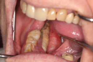 But the femur isn’t the only area affected by osteoporosis drugs. The other vulnerable area in the body is the alveolar bone in the mouth, which turns over at a rate 10 times faster than that of long bones. This is why drug-induced osteonecrosis of the jaws (DIONJ) always begins in the alveolar bone.
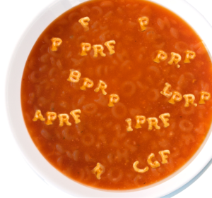 Commercial interests quickly co-opted the conversation, drowning out the voices of clinicians and researchers like Drs Arun Garg and Robert E. Marx—the co-discoverers of PRP—who did not want to see this low-cost technology exploited by profit-hungry manufacturers of centrifuge devices. There was also an enormous amount of misinformation being promulgated by certain medical/dental experts who recognized the enormous therapeutic potential of autologous growth factors and seized the opportunity to establish a name for themselves in the scientific community. The medical literature became saturated with articles introducing new terminology to describe slightly modified growth factor compositions, often without much (if any) additional clinical benefit. The result was an alarming lack of standardization in protocols, a nomenclature best described as an alphabet soup of acronyms, and an overwhelming sense of confusion among clinicians. That’s why Dr Arun Garg published the first edition of this book back in 2015, to set the record straight.
 Above all, he wanted to refocus the conversation about platelet-derived therapies in order to make PRP accessible again to the practicing clinician. He purposefully titled the book Autologous Blood Concentrates instead of PRP or PRF as a way to signal his desire to focus on the science and not the politics swirling around it. He wanted the book to reach clinicians regardless of which machine or nomenclature they were most familiar with. For Dr Garg, it was all about making this low-cost technology accessible and useful to clinicians to better their patients’ outcomes and lives. Everything else was secondary.
 Arun K. Garg, DMD, served as a full-time professor of surgery and director of residency training in the Division of Oral and Maxillofacial Surgery at the University of Miami School of Medicine for nearly 20 years. He has authored more than 8 textbooks and over 150 scientific journal articles and trained thousands of dentists and dental specialists over the course of his career. He is the founder of Implant Seminars, a leading dental education company, and the Garg Foundation in the Dominican Republic, which offers state-of-the-art dental treatment to the community free of charge. In addition, he maintains several private practices throughout South Florida.

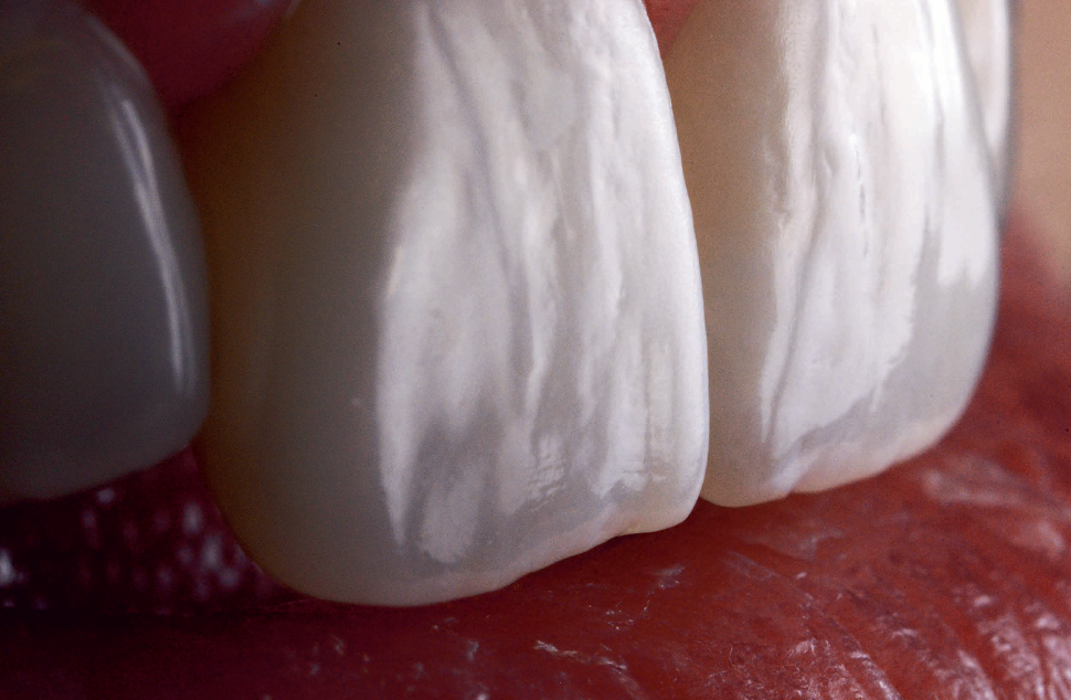
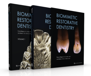
 Yet biomimetics is still met with some skepticism because of a lack of belief in adhesion, despite scores of evidence to the contrary. Dentists across the world are unnecessarily sacrificing tooth structure, and as Dr Magne points out, “the same clinicians are seduced by manufacturers with products that may be easier and faster to use but not better.” That’s why Dr Magne and Dr Belser have updated their book to include all the research and techniques developed in the last two decades. Much has changed, with the development of digitally guided implant dentistry, guided tissue regeneration, and CAD/CAM restorations, yet the principles of biomimetics have not. These are just new tools, not new concepts.
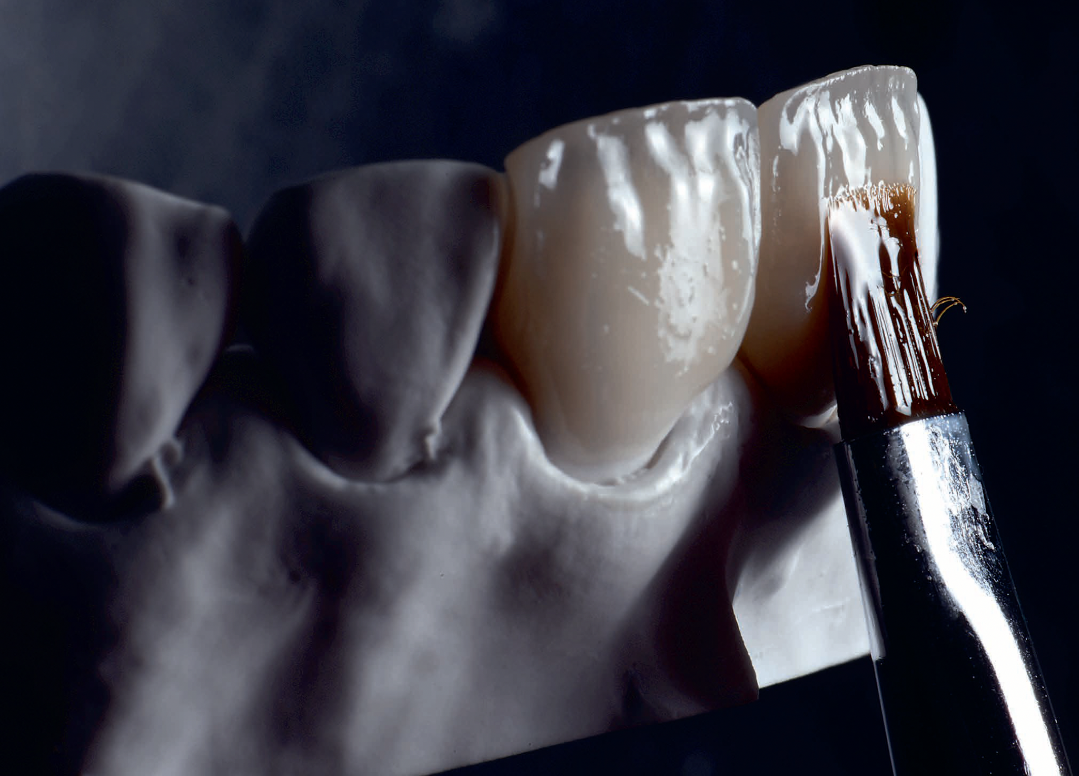
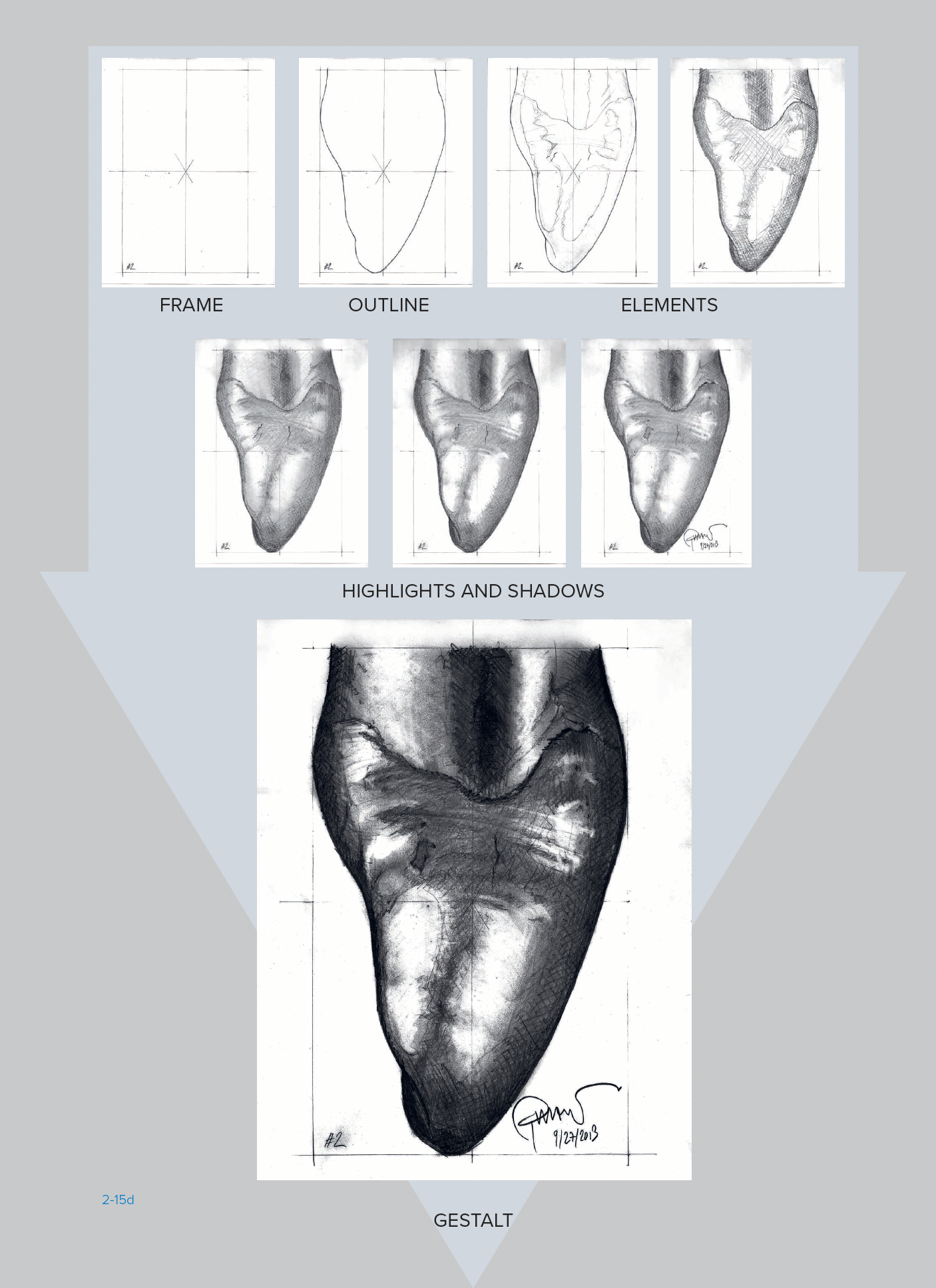
 Dr Pascal Magne is an Associate Professor with tenure and the Don and Sybil Harrington Foundation Professor of Esthetic Dentistry in the Division of Restorative Sciences at the University of Southern California Herman Ostrow School of Dentistry in Los Angeles. He graduated from the University of Geneva Dental School in Switzerland in 1989 with a Med Dent degree and later obtained his doctorate in 1992 and his Privat Docent degree in 2002. Dr Magne received postgraduate training in fixed prosthodontics and occlusion, operative dentistry, and endodontics and was a lecturer at the same university beginning in 1989 until 1997. From 1997 to 1999, he was a Visiting Associate Professor at the Minnesota Dental Research Center for Biomaterials and Biomechanics at the University of Minnesota School of Dentistry. After concluding 2 years of research, Dr Magne returned to the University of Geneva Dental School before being recruited to the University of Southern California in February 2004. He is the recipient of multiple awards and the author of numerous clinical and research articles on esthetics and adhesive dentistry, as well as an internationally known mentor and lecturer on these topics. The first edition of this textbook has been translated into 12 languages and is considered one of the most outstanding books in the field of adhesive and esthetic dentistry. Dr Magne is a founding member of the Academy of Biomimetic Dentistry and a mentor of the Bio- Emulation think-tank group. In 2012, he launched a revolutionary approach to the teaching of dental morphology, function, and esthetics (the 2D/3D/4D approach) for freshman students at the Herman Ostrow School of Dentistry at USC.